Cellular-, 3D- and Correlative Electron Microscopy

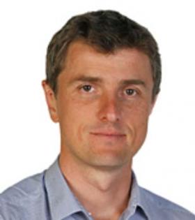
Dr. Yannick Schwab
Team Leader & Head, Electron Microscopy Core Facility
EMBL, Heidelberg, Germany
EMBL Australia and the Cell Motility & Mechanobiology group are proud to host Dr. Yannick
Schwab for a special seminar at 1:30-2:30pm, Thursday 20th July 2017.
Cellular-, 3D- and Correlative Electron Microscopy
Abstract Correlative light and electron microscopy (CLEM) is a set of techniques that allow
data acquisition with both imaging modalities on a single object. One common challenge when
trying to combine imaging modalities on the same sample is to identify space cues (external or
internal) to track single objects when switching from light microscopy (LM) to electron
microscopy (EM). On adherent cultured cells, we have previously developed specific substrates
with coordinates to precisely record the position of cells (Spiegelhalter et al., 2009).
On more complex specimens, such as multicellular organisms, this targeting is even more
critical, as systematic EM acquisition of their entire volume is close to impossible. For this
reason, we are developing new methods to map the region of interest (ROI) within large living
specimens, taking advantage of structural hallmarks in the sample that are visible with both LM
and EM. The position of the ROI is mapped in 3D by confocal or multiphoton microscopy and
then tracked at the EM level by targeted ultramicrotomy (Kolotuev et al. 2009; 2012; Goetz et al.
2014). Relying on structural features of the sample as anchor points, the cell or structure of
interest can then be retrieved with sub-micrometric precision (Durdu et al. 2014, Goginashvili et
al. 2015, Hampoelz et al 2016).
References
• Spiegelhalter C et al. (2010), PLoS ONE 5(2):e9014. doi: 10.1371/journal.pone.0009014
• Kolotuev I, Schwab Y, Labouesse M. (2010), Biol. Cell 102(2):121-132. doi: 10.1042/bc20090096
• Kolotuev I… Schwab Y. (2012), Methods Cell Biol. 111:203-222. doi: 10.1016/b978-0-12-416026-2.00011-x
• Goetz JG, et al. (2014), Cell Rep 6(5):799-808. doi: 10.1016/j.celrep.2014.01.032
• Durdu S, et al. (2014), Nature 515(7525):120-124. doi: 10.1038/nature13852
• Goginashvili A, et al. (2015), Science 347(6224):878-882. doi: 10.1126/science.aaa2628
• Hampoelz B et al. (2016), Cell 166(3):664-678. doi: 10.1016/j.cell.2016.06.015
Back to top
Archive
- February
- March
- April
- May
- June
- July
- August
- September
- The complex lives of glia after...
- Randwick Precinct Initiating Early...
- Randwick Precinct Cancer Genomics...
- What are the mechanisms behind...
- Congratulations to new Associate...
- Medicine Learning L&T Forum
- Revolution in Breast Cancer -...
- The Split Personality of Glutamate...
- Lowy Cancer Research Centre...
- Cancer Metabolism roundtable
- Single Cell Epigenomics for...
- October
- November
